Blizard Institute - Faculty of Medicine and Dentistry

CREATE Lab

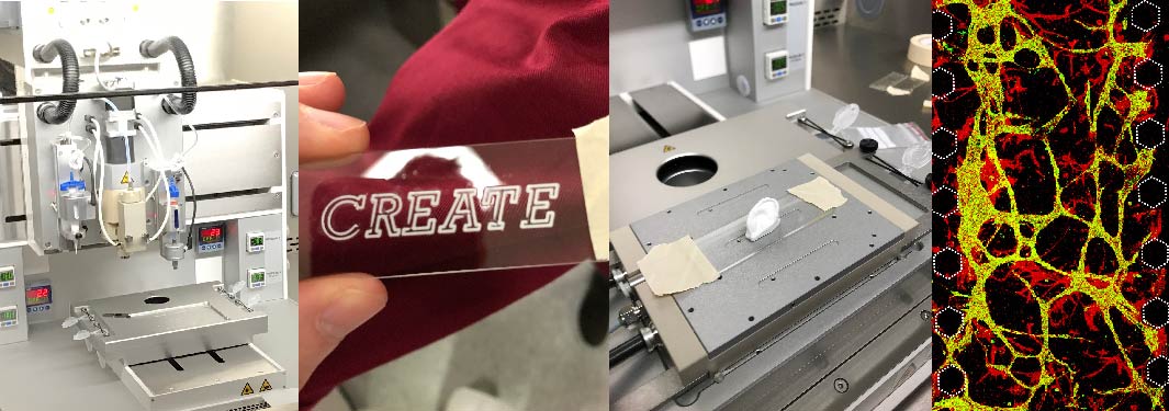
Various pieces of equipment in the CREATE Lab

The CRoss-InstitutE Advanced Tissue Engineering (CREATE) Lab is an established core facility dedicated to biofabrication of advanced 3D tissues and microfluidic devices. It houses state-of-the-art equipment for 3D bio-printing, microfabrication, and device analysis. The aim of the CREATE Lab is to support the development of next-generation 3D tissues and disease models for use in biomedical research and regenerative medicine.

As a cross-faculty initiative, the CREATE Lab was established through the Queen Mary Strategic Facilities Investment Fund and support from the Barts Charity. It is open to all Queen Mary researchers and external partners, with a view to promoting cutting-edge interdisciplinary research.

News

CREATE lab Twitter feed now active! Find out the latest news in the world of Biofabrication @QMCREATE.

The CREATE Lab participated in community engagement by showcasing organ-on-chip technology during the fifth Festival of Communities. We were joined by more than 5,600 local residents in Stepney Green Park, and by more than 2,400 visitors on the Sunday at Queen Mary’s Mile End campus. Find out more.

3D Discovery bio-printing system (regenHU)

The 3D Discovery Biosafety system is an advanced 3D printer enclosed within a class II biosafety cabinet. The system can be configured with up to 4 print heads, including syringe-based extrusion and micro-valve droplet dispensing. The system also provides temperature control for print heads and the printing platform, as well as a UV tool for photo-crosslinking and curing. Printer operation and construct design is controlled through the external computer and CAD/CAM software.

Micro-fabrication suite

The CREATE Lab houses a suite of equipment for photolithography and fabrication of microfluidic devices and micropatterned surfaces. Key pieces of equipment include a spin coater (SPIN150i, SPS), UV masking system (KUB-6, Kloe), hot plates, plasma oxidiser (Zepto, Henniker), and a thermoevaporator (Moorfield nanoPVD). In addition, the lab provides an optical profilometer (Profilm 3D, Filmetrics) for characterisation of microfabricated surfaces, and syringe pumps for introduction of dynamic fluid flow.


Filament based 3D printer Craftbot2

This equipment is suitable for printing PLA and ABS to produce custom sample holders/parts. The facility offers support in CAD drafting.

Training

Facility staff will provide all necessary training and technical support for users to operate the 3D printing and microfabrication equipment.  This includes formal training sessions, one-on-one support, and training manuals and documentation.


Project development

The CREATE Lab team can also assist with new project development, including biomaterial selection, construct design, and optimisation of biofabrication processes.  In addition, we can advise and consult on the technical aspects and costings for research proposals involving biofabrication technologies.


Cell culture facilities

The CREATE Lab includes dedicated cell culture facilities for users to maintain and propagate cells to be used in their 3D bio-printing and microfluidic applications.

All new users are encouraged to first discuss project plans with either the Academic Lead or Research Engineer for the CREATE Lab prior to commencement of new research projects.

To access the CREATE Lab facilities, users must complete a health and safety induction for the Blizard Institute and receive formal training from the lab’s research engineer.  Users will only be allowed to operate the equipment independently when they have satisfactorily completed the training programme.

Equipment is bookable through an on-line booking system.  Users will be charged for usage of standard consumables (syringes, needles, cell culture supplies), while specialised biomaterials or supplies should be provided by the users themselves.  

Academic Lead and Director, Professor John Connelly, Professor of Bioengineering, j.connelly@qmul.ac.uk

Biofabrication Research Engineer, Dr Liisa Blowes, l.m.blowes@qmul.ac.uk 

CREATE Key Publications

  • Sadler E, Stephen AS, Allaker RP, Crick CR. Engineering Biofouling Resistant Materials Through the Systematic Adaptation of Surface Morphology. Adv. Mater. Interfaces 2023, 2202532. https://doi.org/10.1002/admi.202202532
  • Button R, Harwood C, O’Shaughnessy RFL. Phosphoproteomic analysis of the AKT signalling axis in cutaneous squamous carcinoma progression reveals novel therapeutic targets. bioRxiv 2022.10.03.510591; doi: https://doi.org/10.1101/2022.10.03.510591
  • Jones CFE, Di Cio S, Connelly JT, Gautrot JE. Design of an Integrated Microvascularized Human Skin-on-a-Chip Tissue Equivalent Model. Front Bioeng Biotechnol. 2022 Jul 19;10:915702. doi: 10.3389/fbioe.2022.915702. PMID: 35928950; PMCID: PMC9343775.
  • Sarmin AM, El Moussaid N, Suntornnond R, Tyler EJ, Kim YH, Di Cio S, Megone WV, Pearce O, Gautrot JE, Dawson J, Connelly JT. Multi-Scale Analysis of the Composition, Structure, and Function of Decellularized Extracellular Matrix for Human Skin and Wound Healing Models. Biomolecules. 2022 Jun 16;12(6):837. doi: 10.3390/biom12060837. PMID: 35740962; PMCID: PMC9221483.
  • Candarlioglu PL, Dal Negro G, Hughes D, Balkwill F, Harris K, Screen H, Morgan H, David R, Beken S, Guenat O, Rowan W, Amour A. Organ-on-a-chip: current gaps and future directions. Biochem Soc Trans. 2022 Apr 29;50(2):665-673. doi: 10.1042/BST20200661. PMID: 35437569; PMCID: PMC9162452.
  • Sarmin AM, Connelly JT. Fabrication of Human Skin Equivalents Using Decellularized Extracellular Matrix. Curr Protoc. 2022 Mar;2(3):e393. doi: 10.1002/cpz1.393. PMID: 35263039; PMCID: PMC9310708.
  • Pundel OJ, Blowes LM, Connelly JT. Extracellular Adhesive Cues Physically Define Nucleolar Structure and Function. Adv Sci (Weinh). 2022 Apr;9(10):e2105545. doi: 10.1002/advs.202105545. Epub 2022 Feb 5. PMID: 35122409; PMCID: PMC8981897.
  • Swiatlowska P, Iskratsch T. Tools for studying and modulating (cardiac muscle) cell mechanics and mechanosensing across the scales. Biophys Rev. 2021 Sep 5;13(5):611-623. doi: 10.1007/s12551-021-00837-2. PMID: 34765044; PMCID: PMC8553672.
  • Connelly JT, Gavara N, Sliogeryte K, Blowes LM. Research Techniques Made Simple: Analysis of Skin Cell and Tissue Mechanics Using Atomic Force Microscopy. J Invest Dermatol. 2021 Aug;141(8):1867-1871.e1. doi: 10.1016/j.jid.2021.02.750. PMID: 34303466.
  • Singh B, Abdelgawad ME, Ali Z, Bailey J, Budyn E, Civita P, Clift MJD, Connelly JT, Constant S, Hittinger M, Kandarova H, Kearns VR, Kiuru T, Kostrzewski T, Kress S, Durban VM, Lehr CM, McMillan H, Metz JK, Monteban V, Movia D, Neto C, Owen C, Paasonen L, Palmer KA, Pilkington GJ, Pilkington K, Prina-Mello A, Roper C, Sheard J, Smith S, Turner JE, Roy I, Tutty MA, Velliou E, Wilkinson JM. Towards More Predictive, Physiological and Animal-free In Vitro Models: Advances in Cell and Tissue Culture 2020 Conference Proceedings. Altern Lab Anim. 2021 May;49(3):93-110. doi: 10.1177/02611929211025006. Epub 2021 Jul 6. PMID: 34225465.
  • Mehanna YA , Sadler E , Upton RL , Kempchinsky AG , Lu Y , Crick CR . The challenges, achievements and applications of submersible superhydrophobic materials. Chem Soc Rev. 2021 Jun 8;50(11):6569-6612. doi: 10.1039/d0cs01056a. PMID: 33889879.
  • Wu Y, Fortunato GM, Okesola BO, Brocchetti FLPD, Suntornnond R, Connelly J, De Maria C, Rodriguez-Cabello JC, Vozzi G, Wang W, Mata A. An interfacial self-assembling bioink for the manufacturing of capillary-like structures with tuneable and anisotropic permeability. Biofabrication. 2021 Apr 8;13(3). doi: 10.1088/1758-5090/abe4c3. PMID: 33561850.
  • Costa P, Blowes LM, Laly AC, Connelly JT. Regulation of collective cell polarity and migration using dynamically adhesive micropatterned substrates. Acta Biomater. 2021 May;126:291-300. doi: 10.1016/j.actbio.2021.03.032. Epub 2021 Mar 16. PMID: 33741539.
  • Laly AC, Sliogeryte K, Pundel OJ, Ross R, Keeling MC, Avisetti D, Waseem A, Gavara N, Connelly JT. The keratin network of intermediate filaments regulates keratinocyte rigidity sensing and nuclear mechanotransduction. Sci Adv. 2021 Jan 27;7(5):eabd6187. doi: 10.1126/sciadv.abd6187. PMID: 33571121; PMCID: PMC7840118.
  • Almeida FV, Gammon L, Laly AC, Pundel OJ, Bishop CL, Connelly JT. High-Content Analysis of Cell Migration Dynamics within a Micropatterned Screening Platform. Adv Biosyst. 2019 Aug;3(8):e1900011. doi: 10.1002/adbi.201900011. Epub 2019 Jun 13. PMID: 32648701.
  • Kenny FN, Drymoussi Z, Delaine-Smith R, Kao AP, Laly AC, Knight MM, Philpott MP, Connelly JT. Tissue stiffening promotes keratinocyte proliferation through activation of epidermal growth factor signaling. J Cell Sci. 2018 May 16;131(10):jcs215780. doi: 10.1242/jcs.215780. PMID: 29669739.
Back to top