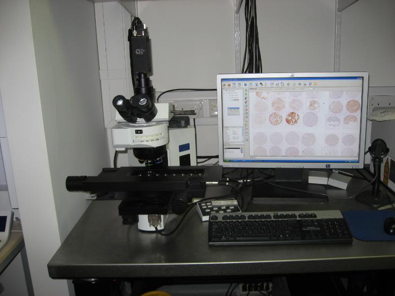
Leica Ariol System for Automated Imaging of Immunohistochemistry Slides

The Ariol system is used for the scanning and quantification of immunohistochemical staining and immunofluorescence. The main application is the collection and analysis of data from tissue sections and tissue microarrays including those stained by fluorescence in situ hybridisation. Nuclear, cytoplasmic and membranous markers can be quantified in terms of intensity, size and number of positive events.
Olympus BX61 microscope with Prior stage to accommodate eight slides. X-Cite 120 Q light source anf filter sets for DAPI, Spectrum Aqua, Spectrum Green, Spectrum Orange and Far Red. Has 1.25x and 5x objectives for locating sample and 10x, 20x, 40x and 60x objectives for image acquisition. Olympus iAi camera and dedicated Ariol software.
Costs
Price per hour : £12.5
Price O/N :
Contact details
Facility : BCI Microscopy Facility
Campus : BCI Charterhouse Square
Address : 3nd Floor, John Vance Science Centre, Charterhouse Square, London EC1M 6BQ
Manager : Linda Hammond
Contact : 020 7882 5132