Advanced Molecular Imaging Service (AMIS)
AMIS, established in 2016, has been supported with over £2.2m of strategic investment to draw together more than 81 pieces of state-of-the-art advanced molecular imaging equipment.
Search
-
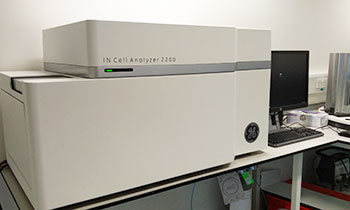
INCA 2200
Super-fast, sensitive, and flexible wide-field cell imaging system for a broad range of high-content assays. The 2200 is the only HCA system on the market that offers deconvolution image restoration resulting in more accurate image segmentation and quantification. Objectives fitted: 4x, 10x, 20x and 40x. Filter set: DAPI/FITC/Cy3/Cy5 and Transmitted light. sCMOS Camera
-
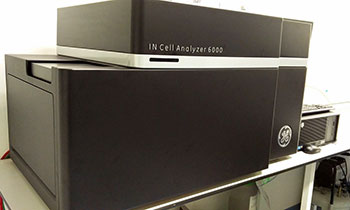
INCA 6000
IN Cell Analyzer 6000 is a highly sensitive, laser-based confocal imaging platform for the most demanding high-content assays and screens. Featuring a novel and proprietary optical system that incorporates an iris-like variable aperture design and next-generation sCMOS technology, IN Cell Analyzer 6000 enables you to optimize for speed and image quality for challenging and variable assays.
-

GE INCarta
GE new dedicated high content analysis software allowing bespoke analysis protocols to be written removing the bottleneck of image analysis
-
GE Developer ToolBox
Dedicated high content analysis software which is user definable allowing your analysis to be automated and unbias. We currently have 4 analysis machines for users.
-

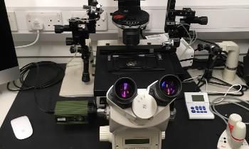
Olympus iX81 Epifluorescence Microscope
This Olympus iX81 is an inverted, fully motorised and environment-controlled microscope for brightfield, phase contrast and epifluorescence imaging applications, suited to live and fixed samples on slides, 35 mm dishes or multi-well plates.
-
Leica SP5 Confocal
An upright laser scanning confocal microscope equipped for high temporal and spatial resolution imaging of fixed cells or tissue sections, as well as live tissue and in vivo intravital techniques.
-

Leica SP8 Confocal
An upright laser scanning confocal microscope equipped for high temporal and spatial resolution imaging of fixed cells or tissue sections, as well as live tissue and in vivo intravital techniques.
-
-800x552.jpg
)
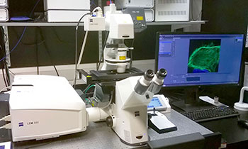
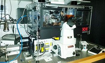
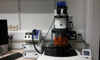
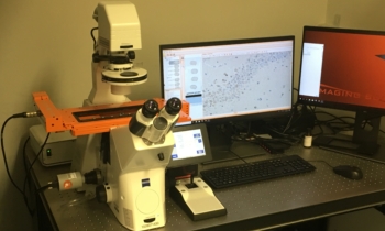
Zeiss LSM 800 Confocal
This inverted Zeiss LSM 800 is a fully motorised laser scanning confocal microscope with a fast piezo Z-drive and environment control (temperature and CO2). It is ideally suited to extended live cell imaging, as well as high-resolution fixed sample analyses.
-
.jpg
)
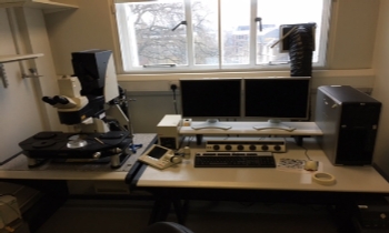
Zeiss Axioscop epifluorescence microscope
This upright microscope enables brightfield, DIC and fluorescence imaging applications with fixed and live samples. The system is ideally configured for intravital imaging of transparent tissues (e.g. cremaster muscles, mesentery).
-
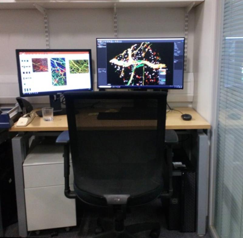
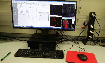
Image analysis workstations
Three high-powered analysis workstations available for remote and in-person use, running dedicated image visualisation and analysis software packages. Available software includes the Imaris 3/4D image visualisation and analysis package, Fiji (ImageJ), Matlab, Anaconda and ZEN. The CMR Advanced Bio-imaging Facility can provide image analysis guidance and support to facility users.
-

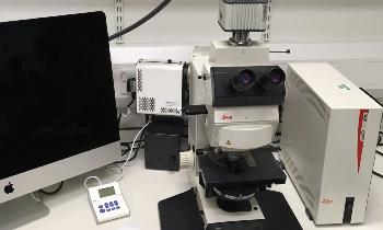
Leica SP8 DIVE Multiphoton with FALCON FLIM
A state-of-the-art upright multiphoton microscope with combined laser scanning confocal and high-speed fluorescence lifetime imaging capability. With a large stage area, long working distance lenses and upright configuration, this microscope is ideally suited to diverse sample types that may include standard slide mounts and 35 mm dishes, as well as custom-designed mounts for more complex sample types and intravital experiments.
-
Zeiss LSM 880 Laser Scanning Confocal Microscope (inverted) with Fast Airyscan module
State-of-the art, fast scanning, near super-resolution confocal microscope. The Airyscan detector makes it possible to increase the resolution of your imaging far beyond the resolution of a classic confocal point scanning microscope. In addition, images can be acquired with high sensitivity and high speed.

This core confocal microscope facility was made possible by a grant from Barts Charity
-

Siemens Inveon microPET/CT
This is a nuclear imaging modality that requires the injection of radioactivity in order to acquire data. Positron-emitting radionuclides such as F-18, Ga-68, Cu-64 and Zr-89 are used.
microCT Scanning: This instrument can also be used as a microCT and the maximum resolution is about 20 microns. The instrument is not that easy to use unassisted so you will require training. Please fill in the training request form under the Request Services tab. Alternatively, contact us under the Request Services, Imaging Request tab if you would like us to carry out a study for you. -
IVIS Lumina III Fluorescence and Bioluminescence camera
The IVIS Lumina III is capable of imaging both fluorescent and bioluminescent reporters. The system is equipped with up to 26 filter sets that can be used to image reporters that emit from green to near-infrared. Superior spectral unmixing can be achieved by Lumina III’s optional high resolution short cut off filters.
-
Visual Sonics Vevo 2100 Ultrasound Instrument
Ultra High-Frequency Imaging Platform with Linear Array Technology. This Ultrasound is capable of 3D-Mode Imaging & Volume Analysis, Nonlinear Contrast Imaging and blood flow quantification via Power Doppler. Two different Transducers are available. Its very operator dependent so you will require training and practice.
-

Leica TCS SP5 laser scanning confocal microscope with two photon capability
Leica TCS SP5 confocal microscope system is equipped with a number of lasers for probe excitation between 355 nm and 633 nm.A steplessly tunable detector system can record any wavelength emitted from the sample and up to five channels may be recorded simultaneously.Two different scanners are available, a conventional scanner for high quality imaging and a fast resonant scanner for applications such as calcium imaging in living tissue. Two photon SpectraPhysics Mai Tai laser is suitable for imaging of living tissues with greater depth penetration.
-

Leica DMRA2 microscope/QIClick colour camera
Digital colour camera/ upright microscope system suitable for bright field colour microscopy, also capable of fluorescent imaging.
-
Leica DMRA2 microscope/Hamamatsu Orca ER camera
Digital camera/ upright microscope system suitable for epifluorescent microscopy.
-
Leica DMIRE2 microscope/Q\Click mono camera
Digital camera/inverted microscope system suitable for epifluorescent microscopy and brightfield microscopy.
-

Amnis ImageStream X MkII
Combining flow cytometry with microscopy, the ImageStream X Mark II generates standard flow cytometry data, plus fluorescence microscopy and brightfield images for every cell
-
Leica DM4000 Epi-Fluorescence Microscope (upright)
The Leica epi-fluorescence microscope allow basic four colour wide-field image capture of fluorescent and histology samples with a monochrome CCD camera.
-
Leica DM5000B Epi-Fluorescence Microscope (upright)
The Leica epi-fluorescence microscope allow basic four colour wide-field image capture of fluorescent and pathology stained samples via monochrome and colour CCD cameras.
-
Zeiss Axiovert 200M TimeLapse Epi-fluorescent Microscope (inverted) at BCI
The Zeiss epi-fluorescence microscope combined a Solent Scientific CO2 and temperature contolled chamber allow fluorescent, phase contrast and DIC imaging of living samples
-
Olympus IX70 TimeLapse Epi-Fluorescent Microscope (inverted)
The Olypus IX70 is equiped with a Cairn Scientific Optosplit allowing true simultaneous imaging of 2 channels wich is ideal for Calcium Imaging
-
Zeiss 710 Laser Scanning Confocal Microscope (upright)
The LSM 710 is an upright confocal point scanning microscope system with a number of lasers for probe excitation between 405 nm and 633 nm. It is equiped with a 32 channel spectral detection unit and can be used for multipoint, tile and timelapse imaging
-
Zeiss 880 Laser Scanning Confocal Microscope With Fast Airyscan and Multiphoton (inverted)
The Zeiss LSM 880 is equiped with a full range of laser lines and spectral detetion GaAsP unit to cater for point scanning needs. Additionally it has a Fast Airyscan module allowing superresolution imagaing at speeds up to 30 frames per second. An additional femtosecond laser and NND Big2 unit permits multiphoton imaging.
-
Spinning Disk Confocal Microscope (inverted)
The spinning disk system is available for fast dynamic live cell imaging and also fast collection of 3D datasets.
-
Zeiss PALM Microbeam Laser Microdissection Microscope at Blizard
The Zeiss PALM system is designed to allow dissection of the biomarkers and cells from Tissue.
-
Nikon Eclipse 80i Stereology Microscope (upright)
The Stereology Microscope can take very lage overview scans in Brightfield and Fluorescence Microscopy
-
Leica Dissection Microscope
Sample preparation and imaging of samples where stereo vision is required
-
X-Clarity Tissue Clearing Cooled Electrophoresis
Cooled electrophoresis machine to clear tissue embedded in an acrylamide matrix. Allows imaging of thick specimens like whole mouse brains and spinal chords.
-
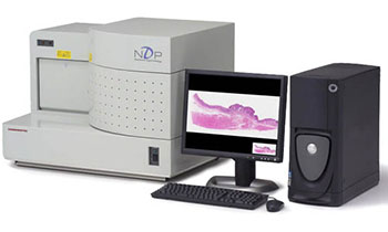
Hammatsu NanoZoomer 2.0-HT Digital slide scanner: C9600
2D image analysis. 210 slide capacity for light microscopy scanning. Fluorescence scanner 20 slide capacity.
-
.jpg
)
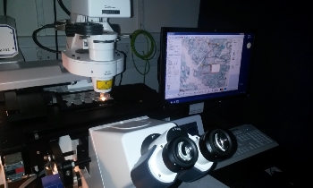
Zeiss 710 Laser Scanning Confocal MIcroscope (inverted)
The LSM 710 confocal microscope is equipped with four lasers and a 34 channel spectral system. The microscope is inverted and fitted with an environmental chamber, allowing users to perform live imaging experiments that can be recorded as a time series.
-
.jpg
)
Zeiss 510 Laser Scanning Confocal MIcroscope (inverted)
The LSM 510 confocal microscope is inverted and equipped with four lasers.
-
Nikon Spinning Disk Confocal Microscope (inverted)
The Nikon spinning disk confocal microscope is suitable for imaging of dynamic processes in living cells and fast acquisition of stacks in fixed preparations. The spinning disk ensures the sample is illuminated and light detected at multiple points simultaneously.The system is equipped with 6 lasers. Two sCMOS cameras allow imaging of both confocal and widefield applications. The microscope has an environmental chamber for live imaging and the piezo stage maintains focus throughout time lapse acquisitions.
-
Zeiss PALM Microbeam Laser Microdissection Microscope at BCI
The P.A.L.M. Microbeam system is for the microdissection and capture of specified regions from tissue samples for subsequent extraction of DNA, RNA or protein.The system works by a process of laser dissection, followed by pressure catapulting. Samples can be collected from paraffin-embedded sections, frozen sections or cultured cells. Both brightfield and fluorescence light sources.
-

Zeiss Axiovert 200M TimeLapse Epi-fluorescent Microscope (inverted) at BCI
This inverted, epi-fluorescence microscope with environmental chamber allows fluorescence and phase contrast imaging of live cells. The main application for this microscope is the sequential imaging of multiple, pre-defined locations in a culture plate over a period of time using the Mark & Find module of AxioVision software.
-

DeltaVision Live Imaging System
The DeltaVision system has an inverted research microscope with environmental chamber and fast, high resolution camera. It is suitable for imaging cells on slides or in culture dishes. Fluorescence, brightfield, phase contrast and DIC. Allows multi-position time lapse imagining of Z stacks with high accuracy. The main application is imaging cell division in detail.
-

IncuCyte Live Imaging System
The IncuCyte ZOOM Imaging system allows simultaneous imaging of up to six culture vessels within a tissue incubator. Image data is collected and processed by a networked external hard drive that generates time based growth curves. Phase and fluorescence measurements.
-
Zeiss SteREO Lumar V12 stereomicroscope
This stereo microscope is used for 3D observation and imaging of small objects. Both brightfield and fluorescence options available.
-
.jpg
)
Zeiss Axioplan Epi-fluorescence Microscope (upright)
An upright, epi-fluorescence microscope for brightfield and fluorescence imaging of tissue sections and cells.
-
.jpg
)
Zeiss Axiophot Epi-fluorescence Microscope (upright)
An upright, epi-fluorescence microscope for brightfield and fluorescence imaging of immunohistochemistry samples.
-

Leica Ariol System for Automated Imaging of Immunohistochemistry Slides
The Ariol system is used for the scanning and quantification of immunohistochemical staining and immunofluorescence. The main application is the collection and analysis of data from tissue sections and tissue microarrays including those stained by fluorescence in situ hybridisation. Nuclear, cytoplasmic and membranous markers can be quantified in terms of intensity, size and number of positive events.
-

3D Histech Pannoramic 250 High Throughput Digital Slide Scanner
The Pannoramic 250 is a high throughput, brightfield scanner with a 250 slide capacity. Allows brightfield scanning of whole tissue sections or TMAs and subsequent analysis using Densito Quant, Nuclear Quant and Membrane Quant modules.
-
NanoZoomer S60 fluorescence and brightfield slide scanner
The NanoZoomer S60 slide scanner from Hamamatsu has the capacity for 60 fluorescence or 60 brightfield slides.
-
NanoZoomer S210 high throughput brightfield slide scanner
The NanoZoomer S210 from Hamamatsu has the capacity for 210 brightfield slides.
-
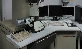
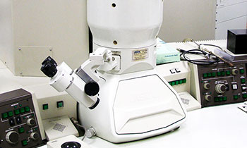
Jeol JEM 1230 TEM
Transmission Electron Microscope mainly operated at 80 kV but can operate up to 120kV. It is equipped with Morada CCD camera and iTEM software.
-

PELCO easiGlow™ Glow Discharge Cleaning System
This device generates a plasma under vacuum. Is used to clean carbon-coated TEM grids which otherwise have the tendency to be hydrophobic. Glow discharge treatment with air will make film surfaces negatively charged and hydrophilic and allow the easy spread of aqueous solutions.
-
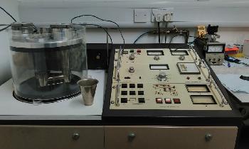
Polaron Freeze Fracture equipment
The Freeze Fracture equipment consist in a vacuum coating unit operating at 10-6 torr with a Platinum and a carbon evaporating source.
-
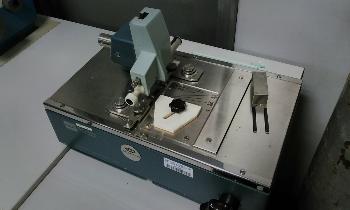
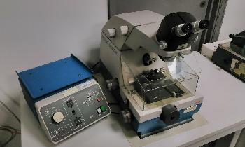
Glass Knifemaker for Reichert Jung Ultracut
Precision glass cutter to make glas knives used for ultrathin sectioning
-
FEI Inspect F
High vacuum FEG source scanning electron microscope (SEM) with EDS and WDS analysis.
EBSD crystallographic texture analysis, grain orientation and mapping
Scanning Transmission Electron Microscopy (STEM) in high-vacuum with nanometre resolution -
FEI Quanta 3D
Scanning electron and ion microscopes are integrated in one system. Can be used as FEG Environmental scanning electron microscope (ESEM)
Focused ion beam for 5-7nm patterning and platinum deposition. Integrated Gatan cryogenic preparation cold stage
Alto 2500. In situ atomic force microscope within ESEM chamber.
Energy dispersive x-ray analysis (EDS) -
FEI Phenom
Rapid optical and SEM imaging to 20,000x magnification. Specimen to image time 25s. Low voltage operation for uncoated samples
-
Attocube AFM system
Bespoke in-situ AFM for perfoming force measurements on the stage of the FIB-SEM
-
Jeol 2010
Transmission electron microscope operated at 200kV, equiped with EDX detector and Gatan digital camera
-
Gatan Dimple Grinder 356
Used for pre-thinning to near electron transparency to reduce the ion milling times. Produces a final thickness of <3 μm in dimpled specimens
-
Gatan Ultrasonic disk cutter 601
Quickly cut simple holes, unique shapes, or TEM discs from hard, brittle materials such as semiconductors, ceramics, and geological materials
Cut materials ranging in thickness from <40 µm to 5 mm -
Gatan Precision Ion Polishing system
Bench-top system designed to produce high-quality TEM specimens with
exceptionally large, clean, electron-transparent areas.Ion polishing is done by two variable-angle, miniature Penning ion guns. The operating angles of the guns, ± 10. -

Zeiss Super resolution LSM 710 ELYRA PS.1
LSM 710 ELYRA PS.1 is a combination of an inverted laser scanning confocal microscopy 710 (LSM 710) with super resolution imaging microscopy ELYRA PS.1: photo activated localization microscopy (PALM)/Stochastic Optical Resolution Microscopy (STORM) and structured illumination microscopy (SIM). It is suitable for imaging targeting at 10-100nm resolution at single molecule level. In addition, Zen 2012 software, motorised X/Y stage, definite focus, TIRF mode make it capable of acquisition of tiling, multiple positions, time lapse, FRAP and FRET. It has a on stage environment chamber with a 35mm petri dish adaptor linked with a CO2/N2 controller for live cell imaging including hypoxic experiments. Furthermore, it comes with an offline data processing workstation.
-
PerkinElmer Spinning Disk Confocal Micoscope
PerkinElmer UltraView imaging system is an inverted spinning disk confocal microscope with 488/586nm laser lines, with an INTRACEL temperature controller and a Narishige micromanipulator, suitable for fast dynamic fluorescent and bright field live imaging such as organelle trafficking, calcium imaging, chromatin and cell dynamics.
-
Leica DMI4000B Epifuluorescence Microscope
Leica DMI4000B is an inverted epifluorescence widefield (WF) microscope suitable for bright field (BF), phase contrast (PH) acquisition and epifluroscence imaging with DAPI, FITC, TRITC, Texas Red and Cy5 filters
-
Tissuefax
TissueFAXS PLUS is an inversed fluorescence and brightfield system for the scanning and analysis of slides, cytospins, smears and Tissue Microarrays. The system is equipped with TissueQuest/HistoQuest or StrataQuest PLUS image analysis software.
TissueFAXS i PLUS is the most versatile configuration of all TG inverse systems.
It can scan and analyse samples on slides, in microtiter plates in both brightfield and epifluorescence modes. It has filters for Dapi/Cy2/Cy3/Cy5.5/Alexa 595/Cy7.